
Man’s CAD diagnosis complicated by 2 lung conditions: Case report
Patient diagnosed with lung blood clot, organizing pneumonia, and CAD
Written by |
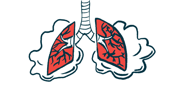
For a man in Japan, cold agglutinin disease (CAD) was complicated by pulmonary thrombosis, which occurs when a blood clot blocks a blood vessel in the lungs, and organizing pneumonia, a rare type of inflammation in the air sacs of the lungs.
The man responded well to treatment with the corticosteroid prednisolone and anti-clotting heparin, leading to near resolution of pulmonary thrombosis and organizing pneumonia within two months.
While it is known that CAD nearly doubles the risk for blood clots, including those that become lodged in the lungs, there have been no reports of organizing pneumonia occurring secondary to CAD.
“This is the first report of [CAD] associated with organizing pneumonia, suggesting a potential link between the two,” researchers wrote in the study “Organizing Pneumonia in a Case of cold Agglutinin Disease with Pulmonary Thrombosis,” published in Internal Medicine.
CAD is a rare autoimmune disease that happens when self-reactive antibodies known as cold agglutinins bind to red blood cells in low temperatures, causing them to clump together and be marked for destruction (hemolysis).
But as these clumps travel through the bloodstream, they can block blood flow in tissues and organs, such as the lungs — potentially causing pulmonary thrombosis.
Organizing pneumonia occurs when buds of inflamed tissue build up in the air sacs in response to damage to the airways. These buds can make it difficult for air to move through the lungs, leading to shortness of breath and chest pain. While uncommon, this rare lung condition can develop secondary to an autoimmune disease.

CAD diagnosis complicated by two other conditions
Now, a team of researchers in Japan described the case of a 46-year-old man whose diagnosis of CAD was complicated by pulmonary thrombosis and organizing pneumonia.
“To our knowledge, there are no other reports of CAD accompanied by PT [pulmonary thrombosis] and OP [organizing pneumonia],” the team wrote.
The man visited the researchers’ hospital after recurrent but self-resolving episodes of chest pain, with the latest being accompanied with shortness of breath. A physical examination revealed jaundice (yellowish skin) and purple-colored spots in both forearms.
Blood testing revealed too few red blood cells, low levels of hemoglobin — the protein in red blood cells that carries oxygen — and high levels of markers of hemolysis. A Coombs direct test, which looks for antibodies bound to red blood cells, came back positive, and further testing confirmed the presence of cold agglutinins in the blood.
These findings suggested the presence of CAD, but the man also showed high numbers of white blood cells and elevated markers of abnormal blood clotting and lung inflammation or damage. However, no signs of cancer or infection were detected.
“Further examinations were performed to enable a precise diagnosis,” the researchers wrote.
Bone marrow analyses ruled out blood cancers, but showed slightly higher counts of B-cells, the type of immune cells that produce antibodies, including cold agglutinins.
A chest X-ray revealed abnormal shadows in both lungs. Chest CT angiography, which uses a special dye to produce pictures of blood vessels, confirmed pulmonary thrombosis in the artery supplying the right lung.
A sample of the lung mucus and a lung biopsy revealed the presence of high numbers of white blood cells, as well as signs of potential tissue scarring and a developing polyp, or growth, within an air sac. However, no bacteria were found in the lung mucus sample.
Based on these findings, the doctors made a diagnosis of CAD complicated by pulmonary thrombosis and organizing pneumonia.
Two months of treatment resulted in near resolution two conditions
The man was started on prednisolone, an immunosuppressive and anti-inflammatory medication, and heparin, which suppresses blood clotting. Treatment resulted in an increase in hemoglobin levels, as well as a reduction in a marker of blood clotting and in abnormal lung shadows.
Heparin was switched to apixaban, another anti-clotting medication branded as Eliquis and also available as generics. After two months, CT scans showed near resolution of both pulmonary thrombosis and organizing pneumonia.
Treatment with Enjaymo (sutimlimab), an injectable antibody approved to treat hemolysis in adults with CAD, was started during follow-up visits.
“The patient’s condition did not worsen within two years of the start of treatment,” the researchers wrote. “Given the similar onset, clinical course, and response to [prednisolone], it was unlikely that both [CAD and organizing pneumonia] merely occurred by coincidence.”
Also, given that pulmonary thrombosis can promote inflammation, it’s possible organizing pneumonia “might have been caused not by CAD but by PT,” the researchers wrote.
But overall, “our findings suggest that CAD may cause not only PT but also OP; therefore, pulmonary lesions should receive particular attention if CAD is encountered,” they concluded.


Leave a comment
Fill in the required fields to post. Your email address will not be published.