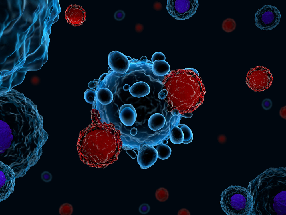
Subset of T-cells Play Role in the Production of Autoantibodies in AIHA, Study Suggests

T follicular helper cells, a subset of T-cells that help other immune cells produce antibodies, seem to play a role in the development of autoimmune hemolytic anemia (AIHA), a mouse study suggests.
The study, “The Role of T Follicular Helper Cells and T Follicular Regulatory Cells in the Pathogenesis of Autoimmune Hemolytic Anemia,” was published in the journal Scientific Reports.
AIHA is an autoimmune disease characterized by the production of autoantibodies that attack and destroy red blood cells. It includes two variants, cold agglutinin disease (CAD) and warm antibody autoimmune hemolytic anemia (wAIHA), depending on whether autoantibodies bind more easily to red blood cells at lower or higher temperatures, respectively.
However, the molecular mechanisms underlying the production of these autoantibodies are still not fully understood, which undermines the design of better therapies.
Previous studies investigating the origin and development of AIHA have focused mainly on immune cells responsible for the production of antibodies (the B-cells) or for controlling the activity of other immune cells (the regulatory T-cells).
In this study, researchers from the Peking University Second Hospital in China and their colleagues decided to explore the potential role T follicular helper cells (TFH) and T follicular regulatory cells (TFR), a subset of regulatory T-cells, in AIHA.
To that end, investigators used a technique called flow cytometry to measure the levels of these cells in the blood of mice with AIHA, as well as to assess some of their properties.
They discovered the number of TFH cells, as well as the ratio between the levels of TFH and TFR cells, were higher in mice that produced large amounts of autoantibodies targeting red blood cells, compared to those that did not produce these harmful antibodies.
Moreover, they found that animals whose blood contained high levels of autoantibodies also had higher levels of interleukin-21 (IL-21) and interleukin-6 (IL-6) — two cytokines that are known to play an important role in the differentiation and function of TFH cells. Cytokines are molecules that mediate immune and inflammatory responses.
They also discovered that mice producing high levels of harmful autoantibodies also had higher expression levels of Bcl-6 and c-Maf — two transcription factors that are known to control the generation, differentiation and function of TFH cells. Transcription factors are proteins that control the activity of certain genes.
In their final experiments, researchers showed that TFH cells isolated from animals that produced large amounts of red blood cell autoantibodies were able to trigger the production of the same autoantibodies when transferred to other animals. These autoantibodies were detected in the second week following cell infusion and remained in high levels for 12 weeks.
“[This study] shed light on the important role of TFH cells in regulating anti-RBC [red blood cell] autoantibody production during (…) AIHA,” the researchers wrote.
“Although the role of the inflammatory environment in the increase in TFH frequency could not be completely excluded, our data strongly suggest that TFH cells participate in B cells differentiation and anti-RBC-antibody production. It is hoped that a greater understanding of TFH and TFR cells can result in promising therapeutic approaches against AIHA,” they added.


