
AIHA Found Linked to Hepatitis E Infection in Rare Case Study
Written by |
The rare case of a man infected with the hepatitis E virus who developed autoimmune hemolytic anemia was described in a study that also highlighted the potential for underdiagnosing autoimmune complications in such infections.
The study, “Unusual Presentation of Hepatitis E with Autoimmune Hemolytic Anemia – A Case Report,” was published in The American Journal of Medicine.
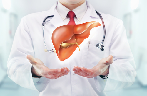
The hepatitis E virus (HEV) causes acute liver injury in 3.5 million people worldwide. The viral infection is also associated with extrahepatic (located or occurring outside the liver) complications including, although rarely, autoimmune hemolytic anemia.
Autoimmune hemolytic anemia (AIHA) comprises a group of rare genetic disorders in which antibodies of the immune system attack and destroy red blood cells. AIHAs are largely grouped into warm or cold types, depending on the ideal temperature for the autoimmune activity. Cold agglutinin disease is the most common type of cold AIHA.
The diagnosis and treatment of HEV-related autoimmune symptoms remains clinically challenging. However, a timely diagnosis is key to prevent further disease development and worse outcomes.
In the study, researchers reported the case of a 65-year-old man who was diagnosed with HEV-related AIHA. He was first examined for a yellowish coloration in the white outer layer of the eye (called sclera) and strong colored urine, which he’d experienced for 20 days. In the previous five days, he had also experienced fever, accompanied by nausea and vomiting. He had no history of bleeding.
On clinical examination, he was pale and jaundiced. His vitals were normal, with no signs of fever. However, clinicians noticed loss of body hair, called alopecia areata and indicative of an autoimmune condition, on the left side of his neck.
Blood lab tests revealed low levels of hemoglobin, the iron-transporting protein that carries oxygen in red blood cells. Moreover, the red blood cells were of unequal size and shape, and the patient had an increased number of immature red blood cells, called reticulocytes.
Blood analysis assessing liver function showed that he had elevated levels of certain liver enzymes and hyperbilirubinemia (an accumulation of bilirubin, which is a yellow molecule that is produced by the breakdown of red blood cells) — all signs of liver damage.
These features led clinicians to suspect that the patient had a form of hemolytic anemia.
Further blood tests showed no signs of blood cancer, and an ANA (antinuclear antibody) test, used to detect autoimmune conditions, was negative. However, the Coomb’s test (which detects antibodies attached to red blood cells) was positive, indicating that the patient had developed AIHA.
More exams showed that he had hepatosplenomegaly (enlargement of the liver and spleen) and tested positive for anti-hepatitis antibodies, indicative of a recent hepatitis E virus infection. No antibodies for hepatitis C or B were found.
“A clinical diagnosis of acute hepatitis E with autoimmune hemolytic anemia was confirmed,” the researchers wrote.
To treat his autoimmune condition, the patient was given steroids, which led to improvements in his hemoglobin levels and liver function test scores.
According to the clinicians, only three cases of hepatitis E-associated AIHA have been reported to date.
“It is possible that different immune alterations as well as the occurrence of AIHA are more frequent in hepatitis E infection than reported till now,” the researchers wrote.
“These patients frequently have been underdiagnosed because their anemia during the course of illness could have been attributed to other causes,” they added.

Leave a comment
Fill in the required fields to post. Your email address will not be published.