Uncommon cellular process in blood may help make AIHA diagnosis
Peripheral erythrophagocytosis found to be characteristic of disease
Written by |


Nearly half of children and teens with autoimmune hemolytic anemia (AIHA) — a group of disorders that includes cold agglutinin disease (CAD) — have signs of erythrophagocytosis, or the removal of old and damaged red blood cells by immune cells, in blood samples, a small study shows.
While erythrophagocytosis typically occurs in the spleen, liver, and bone marrow, its occurrence in circulating, or peripheral, blood is a marker of immune-mediated red blood cell destruction, such as that which occurs in CAD and other forms of AIHA.
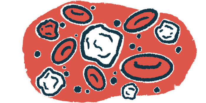
“The recognition of these morphologic findings on the blood smear provides a rapid, yet simple way of making a provisional diagnosis of autoimmune hemolytic anemia universally,” the study’s researcher wrote. A blood smear involves spreading a small amount of blood on a glass slide to examine under a microscope.
The study, “The Prevalence of Peripheral Erythrophagocytosis in Pediatric Immune-Mediated Hemolytic Anemia,” was published in Hematology Reports by a researcher at Mount Elizabeth Hospital in Singapore.
In AIHA, self-reactive antibodies bind to red blood cells, marking them for destruction in a process known as hemolysis. This results in anemia, where there are too few healthy red blood cells to carry oxygen in the body. The disease can be classified as warm AIHA, CAD, and mixed AIHA, depending on whether the antibodies attach to red blood cells at warm or cold temperatures, or a combination of both. Also, the various forms of AIHA can be called either primary, when its cause is unknown, or secondary, when it arises from underlying illnesses, such as infections and other autoimmune disorders.
“The clinical signs and symptoms of AIHA are often nonspecific and its diagnosis has to rely on laboratory findings,” the researcher wrote, adding that several cellular abnormalities that mark hemolysis “can be seen in the peripheral blood smear.”
The presence of red blood cells inside certain immune cells in circulating blood “appears to be a characteristic [cellular] feature that can distinguish immune-mediated hemolysis from the other causes of hemolytic anemia,” the scientist wrote.

Determining frequency of peripheral erythrophagocytosis in AIHA
While peripheral erythrophagocytosis has been described in several case reports of AIHA, its frequency in this patient population remains unknown.
Here, the researcher retrospectively analyzed the medical records of 12 children and adolescents diagnosed with AIHA at a single hospital from July 2014 too June 2024. The patients’ mean age was 6.7 (range, 9.6 months to 16.6 years) and a third were girls.
Seven patients (58%) were positive on a direct antiglobulin test, also called a direct Coombs test, that checks for antibodies and other immune proteins bound to red blood cells. Three (25%) tested positive for cold agglutinins, the self-reactive antibodies that mark CAD. Two (17%) were positive on both tests. Nearly all the patients (92%) had a condition that predisposed them to AIHA, according to the researcher. These included infections in seven cases (58%), an underlying immune disorder in two cases (16.6%), and medications in two cases (16.6%).
Data on several hemolysis markers showed elevated levels in the blood of more than half of the tested AIHA pediatric patients: bilirubin in six of 11 cases (55%) and lactate dehydrogenase in three of five cases (60%).
All had normoblasts in their bloodstream. Normoblasts are immature red blood cells that are normally present in the bone marrow, but are found in the blood in many conditions marked by anemia.
Blood smear results showed that five pediatric AIHA patients (42%) had peripheral erythrophagocytosis. In comparison, during the same time period, 16 people were diagnosed with hereditary spherocytosis, a condition wherein red blood cells adopt a spherical shape instead of the normal flat disk shape, yet none had erythrophagocytosis.
Differences in the rate of peripheral erythrophagocytosis between the two groups were statistically significant.
“Peripheral erythrophagocytosis is a relatively common and characteristic finding in pediatric autoimmune hemolytic anemia and should be actively looked for in the evaluation of acute hemolysis,” the researcher wrote. “The finding of peripheral erythrophagocytosis may have been under-recognized and the medical personnel responsible for reading blood smears should be adequately trained. A better awareness of the presence of peripheral erythrophagocytosis will improve the diagnostic accuracy and rapidity.”